The Instruments/Entry 5.01/One technique, and what it can and cannot carry
The autoradiograph
The early output was a film, read by eye, and its bands are the image everyone still pictures.
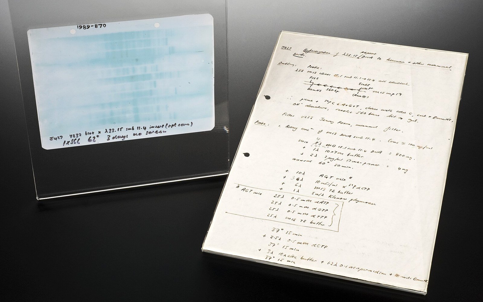
The image everyone pictures when they think of DNA evidence is not what courts use today — it is a piece of X-ray film from a technique already retired.
How the Film Was Made
In the first decade of forensic DNA work, a profile was not a string of numbers. It was a pattern of dark bands on photographic film — an autoradiograph, produced by the controlled decay of a radioactive label. The process began with the Southern blot: DNA was cut with restriction enzymes, the fragments separated by size through a gel, transferred to a nylon membrane, and probed with a short sequence of radioactively labelled DNA designed to bind at a specific location in the genome. The probe's radioactive tag — typically phosphorus-32 — then exposed a sheet of X-ray film laid against the membrane. Where the probe had bound, the film darkened. The result was a ladder of bands, their positions encoding the lengths of the fragments at that locus.
Reading the film required a trained eye and a densitometer. Two samples were compared by asking whether their bands fell at the same position — within a defined tolerance known as a match window, because even identical fragments could run fractionally differently across adjacent lanes of a gel. The judgment was partly visual, partly statistical, and it rested on population databases that, in the mid-1980s, were thin.
From the register
How the image was produced
- Restriction digestion
- DNA is cut at defined sequences by restriction enzymes, producing fragments of different lengths
- Gel electrophoresis
- fragments are driven through a gel by current and separate by size
- Southern transfer
- fragments move from the gel to a membrane, preserving their positions
- Radioactive probing
- a labelled sequence binds to the target locus; its decay exposes photographic film
- Match window
- a defined tolerance used when comparing band positions across gel lanes, because identical fragments can run slightly differently
What the Bands Could and Could Not Say
Alec Jeffreys at the University of Leicester ↗, who developed DNA fingerprinting in 1984, initially worked with multilocus probes — probes that lit up many sites simultaneously, producing a dense, barcode-like pattern that was individually distinctive but hard to handle statistically. Forensic casework quickly moved to single-locus probes, which read one site at a time and allowed a cleaner probabilistic statement: the probability that an unrelated individual chosen at random would share this band pattern at this locus. Run several loci, multiply the probabilities, and the figure became compelling.
The system's weaknesses were real. The method required a relatively large and intact sample: degraded DNA produced faint or absent bands that could not be reliably matched. Mixtures were nearly impossible to interpret — overlapping bands from two contributors merged into ambiguity. Radioactive probes had a working half-life, and the films themselves aged. Interlaboratory reproducibility was a documented concern; the FBI Laboratory ↗ and Home Office-funded programmes both ran proficiency exercises in the late 1980s and early 1990s to establish that different labs reading the same film would reach the same conclusion.
The autoradiograph also carried a persistent risk of misreading. Because the comparison was visual and positional, small differences in gel running conditions could shift a band just enough to create apparent non-matches where there were none, or to create apparent matches where there were none. The match-window approach managed this but did not eliminate it. These limitations mattered: if a band was declared a match on visual inspection alone, the argument in court rested on that inspection being correct.
From the register
Key limitation
- 01Mixture problem — two contributors' bands overlap and cannot be cleanly separated, making mixed samples largely uninterpretable under RFLP
The Image Outlives the Method
Restriction fragment length polymorphism analysis — the technique that produced the autoradiograph — was largely displaced by PCR-based STR methods through the 1990s. PCR required far less sample, tolerated degradation better, automated cleanly and produced a numerical output that computers could handle directly. Capillary electrophoresis replaced the gel slab; a software-generated trace replaced the film.
Yet the autoradiograph has a documentary permanence that digital traces can lack. The films from early cases still exist in evidence archives. When post-conviction review organisations including the Innocence Project began challenging old convictions, those physical films became critical: they could be re-examined with modern reference data or, where the original biological material was preserved, the sample could be re-profiled entirely. The band pattern on a piece of X-ray film is a durable object in a way that a printout from a discontinued instrument is not — which is one reason the transition to digital workflows prompted hard thinking about long-term data preservation that laboratories are still working through.