The Method/Entry 1.05/One technique, and what it can and cannot carry
Capillary electrophoresis
Fragments are separated by size in a thin capillary and detected by fluorescence as they pass a window.
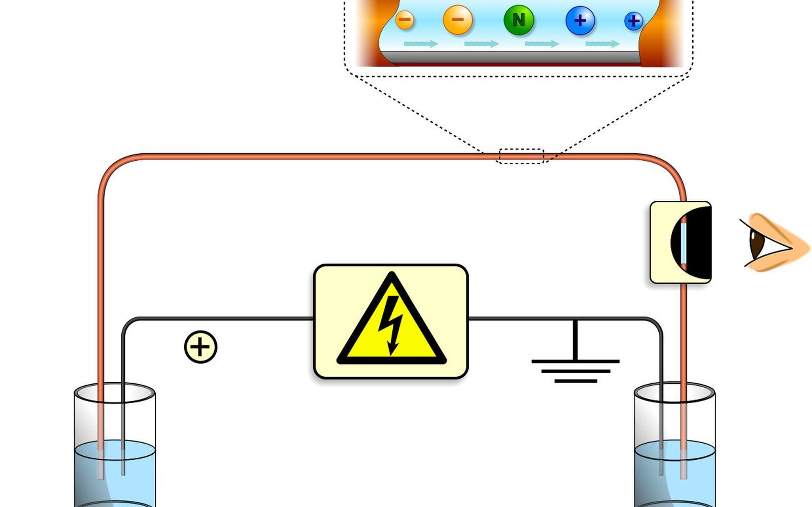
A voltage gradient in a hair-thin tube replaced the gel slab and made modern forensic DNA profiling fast, automated and reproducible.
How the Tube Works
The capillary is a piece of fused-silica tubing, typically around 50 centimetres long and 50 micrometres in internal diameter — roughly the width of a human hair. The tube is filled with a viscous polymer solution that acts as a molecular sieve. A sample containing fluorescently labelled DNA fragments is injected at one end by electrokinetic means: a brief voltage pulse draws a tiny plug of material into the capillary mouth. A sustained voltage — typically several kilovolts — then drives the negatively charged DNA fragments toward the positive electrode, and the polymer matrix slows them according to size. Shorter fragments travel faster; longer ones are retarded more. After a defined run time, each fragment passes a detection window where the polymer coating has been stripped from the silica, and a laser excites the fluorescent dyes attached to the fragments. The resulting emission is captured by a charge-coupled device, and the instrument logs the signal as a function of time, converting time to fragment size by comparison with an internal size standard run in the same capillary in the same injection.
The output is an electropherogram: a series of peaks plotted against base-pair size, with peak height or area indicating the quantity of material. Because STR multiplexes use dyes of different colours to label primer sets, several loci can be read simultaneously in a single capillary run without their signals overlapping. Modern commercial platforms — the Applied Biosystems 3130, 3500 and 3500xL series are those most widely deployed in forensic laboratories worldwide — can run several capillaries in parallel, processing a rack of samples in roughly two hours. That throughput made the instrument a workable partner for national databases that hold millions of profiles.
What the Instrument Sees, and What It Does Not
The fluorescence detector does not know what locus it is reading or whose DNA it is looking at. It records peaks. Everything beyond that — assigning allele designations, calculating a match statistic, deciding whether two profiles are consistent — is interpretation, not instrument output. The distinction matters. National Institute of Standards and Technology ↗ reference materials exist precisely to anchor that interpretation: NIST Standard Reference Material 2391c provides human genomic DNA with certified STR profiles so a laboratory can confirm its instrument is calling alleles correctly before it processes casework samples. Without that kind of external reference, a peak at a given position is just a peak.
The capillary system introduced measurable improvements in precision over the slab-gel methods it replaced. Fragment sizes are determined to within a fraction of a base pair, and the internal size standard corrects for run-to-run variation in temperature and polymer viscosity. Reproducibility across instruments and across laboratories is nonetheless not automatic; it is a product of calibration, maintenance and adherence to validated protocols. SWGDAM ↗ publishes interpretation guidelines that include thresholds for peak height, stutter ratios and the minimum signal required before a peak is treated as a genuine allele rather than noise — all of which are calibrated against the behaviour of a specific instrument on a specific chemistry.
From the register
Key measurements
- Capillary internal diameter
- approximately 50 micrometres, roughly the width of a human hair
- Capillary length
- typically around 50 centimetres for forensic platforms
- Applied voltage
- several kilovolts; drives DNA by charge toward the positive electrode
- Throughput
- 8 or 16 capillaries in parallel; a rack of samples in roughly two hours on modern platforms
- NIST SRM 2391c
- certified human genomic reference material for STR profiling calibration
The capillary does not resolve every ambiguity. Low-template samples produce peaks so small that genuine alleles and background noise occupy the same territory. Mixtures produce peaks from more than one contributor, and the instrument has no way to assign them. Secondary transfer can deposit a contributor's DNA at a concentration so low that only some alleles are detected — a phenomenon called drop-out — while artefacts called stutter peaks appear just below a true allele and can be mistaken for a second contributor's signal. The instrument faithfully records all of it. Whether those signals can be interpreted, and what they establish, is a question that sits downstream of the data and outside the method — one that probabilistic genotyping software increasingly tries to address by modelling peak behaviour statistically rather than calling peaks by eye.
The capillary array succeeded the slab gel in most serious forensic laboratories during the late 1990s and 2000s, and the move was not merely one of speed. Automation reduced the manual handling that introduced contamination; digital data replaced a film read by eye; and instrument logs created an auditable record of every run. Those features did not change what the method can and cannot establish. They changed how reliably the method establishes it.