The Method/Entry 1.01/One technique, and what it can and cannot carry
The Southern blot
The original approach transferred separated fragments to a membrane and probed them, and it needed a large, fresh sample and days of work.
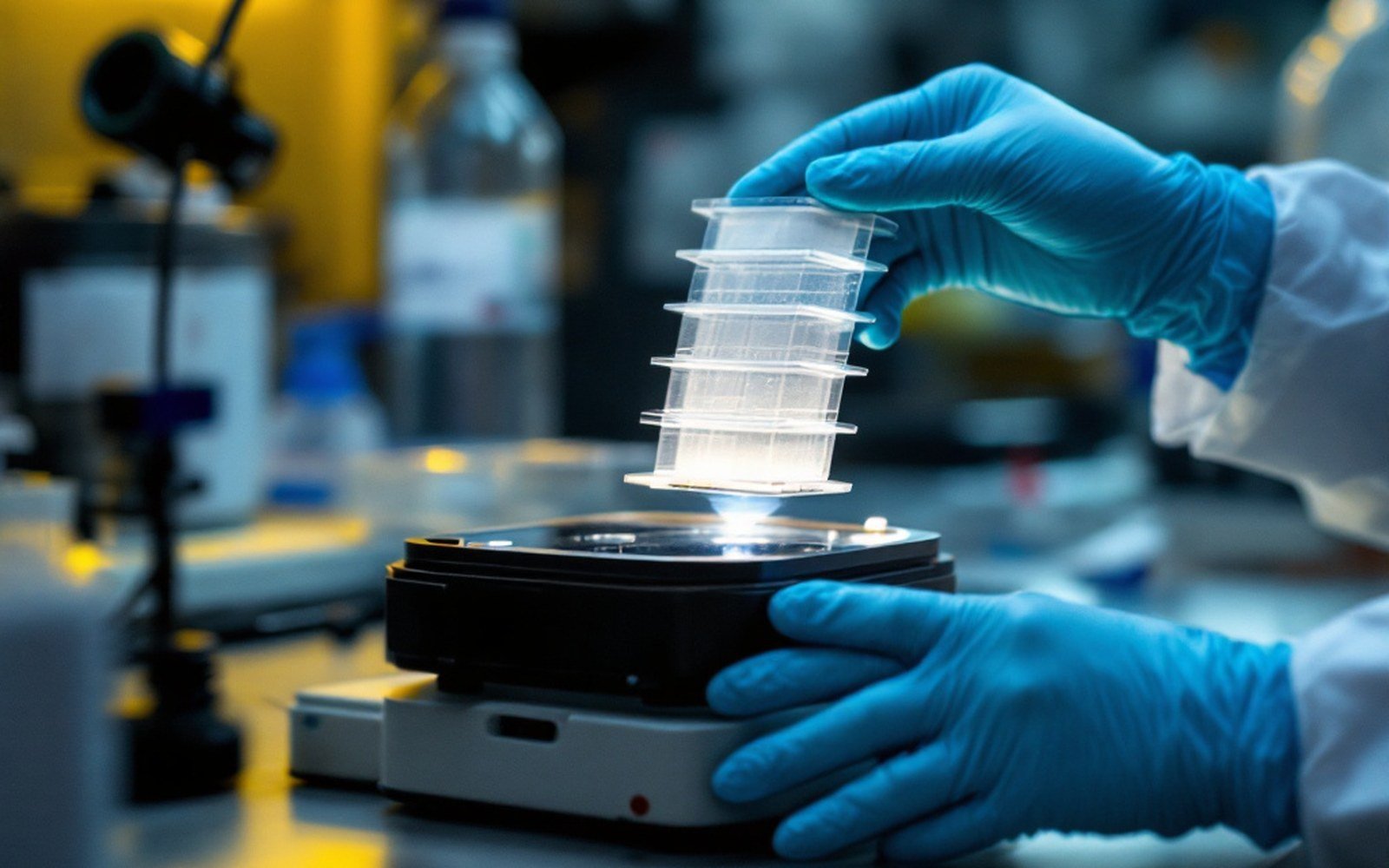
The method that started forensic DNA required a substantial sample, a week of preparation, and a sheet of film read by eye — but it worked, and its logic still runs through every technique that followed.
From a Gel to a Membrane
In 1975, Edwin Southern, working at the University of Edinburgh, published a method for transferring DNA fragments from an agarose gel to a nitrocellulose membrane, where they could be probed with a labelled sequence and detected. The technique bore his name immediately and kept it: the Southern blot. Every derivative that followed — northern blots for RNA, western blots for protein — was named by analogy, a running joke that became standard nomenclature. Southern's original paper described the core logic that forensic scientists would later adopt: separate fragments by size, move them to a surface that holds them permanently, then find the ones you care about with a complementary probe.
The separation step is gel electrophoresis. Digested DNA — cut into fragments at specific sequences by restriction enzymes — is loaded into an agarose gel and driven through it by an electric current. Smaller fragments travel faster and farther; larger ones lag behind. After several hours, the fragments are spread across the gel in an invisible gradient of sizes. The gel itself is fragile, swells in liquid, and cannot be probed directly with any reliability, so the fragments must be moved.
From the register
How the method worked — step by step
- Restriction enzyme digestioncuts genomic DNA at specific sequences into fragments
- Agarose gel electrophoresisseparates fragments by size under electric current
- Capillary transfermoves fragments from the gel to a nylon or nitrocellulose membrane
- Hybridizationradioactively labelled probe binds to complementary target sequences
- AutoradiographyX-ray film exposed by radioactive decay; bands develop at probe-binding positions
- Densitometry and sizingband positions measured against a ladder to determine fragment lengths
Transfer is achieved by capillary action. A stack of absorbent material draws buffer upward through the gel and through a sheet of membrane pressed against it; the DNA fragments migrate with the liquid and bind to the membrane surface. Southern's 1975 procedure used nitrocellulose; nylon membranes came later and proved more durable. The membrane, once dried and baked, carries a permanent replica of the gel's fragment pattern — a fixed, flat record that can be handled, stored and probed.
Probing, Autoradiography, and Reading the Film
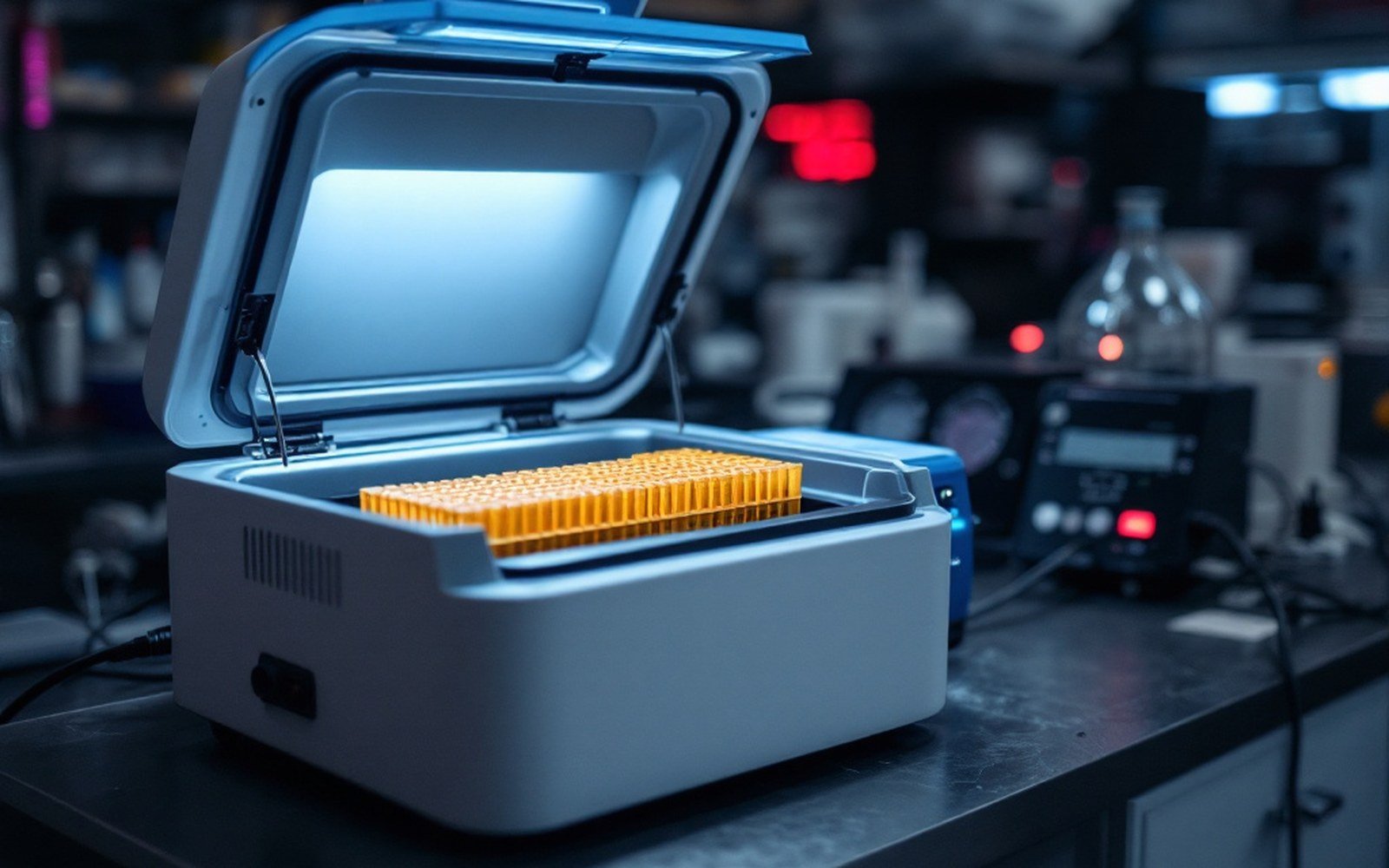
The probe is a short, single-stranded DNA sequence labelled with a radioactive isotope, typically phosphorus-32. Heated to denature the membrane-bound fragments into single strands, then flooded with probe and allowed to cool, the membrane permits the probe to find and bind its complementary sequence — hybridization. After washing away unbound probe, the membrane is pressed against X-ray film in a lightproof cassette. Where probe has bound, the radioactive decay exposes the film. The cassette sits in a freezer for hours or days. When the film is developed, dark bands appear at the positions where the target sequences landed after electrophoresis.
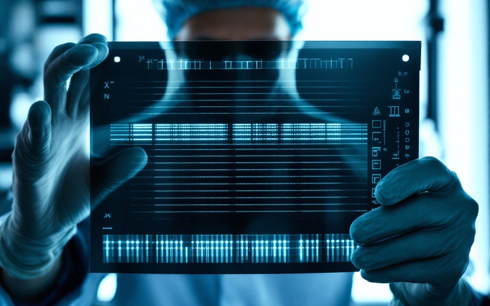
The result is an autoradiograph: a physical sheet of film carrying bands at positions that correspond to fragment lengths, and therefore to the number of repeating units at a given locus. In forensic use, the probe was designed to target a variable region — a VNTR, or variable number tandem repeat — where the fragment length differed substantially between individuals. Two samples run side by side on the same gel, transferred to the same membrane, probed together: if the bands align, the lengths match. If they don't, the samples came from different people.
From the register
What it required vs. what came after
- DNA quantity
- micrograms for Southern blot; nanograms to picograms for PCR-STR
- Time from sample to result
- one to two weeks (Southern blot); one to two days or less (STR/CE)
- Sample condition
- needed intact, high-molecular-weight DNA; degraded samples typically failed
- Output
- physical X-ray film, read by eye with a densitometer; later methods produce a digital electropherogram
- Probe type
- radioactive isotope (phosphorus-32); later methods use fluorescent dyes
Reading the film required a measuring step because the separation is continuous, not digital. Analysts used a densitometer to measure band positions precisely, then compared the measurements against a sizing ladder run in an adjacent lane. A declared match required the band positions to fall within a defined tolerance — the so-called match window — because slight gel distortion and lane-to-lane variation meant that identical fragments rarely landed at exactly the same measured position. The FBI Laboratory and other agencies developed their own match criteria, and the statistical framework for translating a match into a frequency estimate — how common is this fragment length in the relevant population? — was built from databases assembled through the 1980s and into the 1990s.
What the Method Demanded
The Southern blot's requirements were unforgiving. The restriction enzyme needed intact, high-molecular-weight DNA to cut cleanly; degraded DNA produced smearing rather than sharp bands. That meant the sample had to be recent and reasonably well preserved. The quantity needed was substantial — forensic laboratories at the time typically required a visible stain and extracted micrograms of DNA rather than the nanograms or picograms that later PCR-based methods would handle. A hair without its root was useless. A small bloodstain on fabric washed once might yield nothing interpretable.
The timeline from sample to result ran to a week or more, sometimes longer. Restriction digestion, gel electrophoresis, transfer, hybridization, washing, autoradiography and film development were sequential steps with no shortcut. Each required controlled conditions. The radioactive probes had a half-life measured in weeks, so fresh probe had to be prepared regularly. The process was expensive, technically demanding, and concentrated in a small number of specialist laboratories.
Named in this entry
Edwin Southern
Molecular biologist
Described the membrane-transfer technique in 1975 that carries his name, and on which the first profiling method depended entirely.
When Alec Jeffreys at the University of Leicester applied the Southern blot to forensic casework beginning in 1984, he used multilocus probes — sequences that hybridized to many VNTR sites simultaneously — to produce a pattern of many bands, dense enough to be effectively unique to an individual. The result looked like a barcode and was widely described as a DNA fingerprint, a term Jeffreys himself used. That first criminal investigation, in Leicestershire in 1986, used Southern blotting to exonerate an innocent man and then, after a mass screen of thousands of local men, to identify the true perpetrator from a blood sample — establishing every principle that forensic DNA would carry forward.
The National Academy of Sciences and the broader scientific community spent considerable energy in the late 1980s and early 1990s evaluating the Southern blot's statistical foundations, particularly the population databases used to calculate match frequencies. A 1992 National Research Council report ↗ examined these questions directly, recommending both methodological standards and ways to handle uncertainty in the frequency estimates. The debates were sharp, but the underlying method — when properly controlled — withstood them.
Every derivative that followed — northern blots for RNA, western blots for protein — was named by analogy, a running joke that became standard nomenclature.
Why It Was Replaced, and What It Left
PCR-based methods, emerging through the late 1980s after Kary Mullis described the amplification reaction in 1983, needed orders of magnitude less starting material and produced results in hours rather than days. Short tandem repeat analysis, read by capillary electrophoresis, displaced Southern blotting from routine casework almost entirely through the 1990s. By the time SWGDAM published its first STR interpretation guidelines in the early 2000s, the Southern blot was a historical reference point rather than a working tool.
What it left was the conceptual architecture. The idea of separating fragments by size, identifying them at specific loci with probes, comparing profiles between samples, and expressing the result as a frequency in a reference population — all of that came from the Southern blot. The autoradiograph's ↗ bands became the electropherogram's peaks; the match window became probabilistic genotyping software; the film reader became a fluorescence detector. The method changed in every material particular, but the logic that Edwin Southern published in 1975 has never been abandoned.